
 Home
Home
MRI (Magnetic Resonance Imaging) has become an invaluable tool in medical imaging, particularly when it comes to brain health. Understanding MRI brain anatomy is essential for both healthcare professionals and patients, as it provides a clearer picture of the brain’s structure and function. This article takes an in-depth look at MRI brain anatomy, offering key insights into how MRI works, what it reveals, and its role in diagnosing brain-related conditions.
Table of Contents
What is MRI Anatomy?
MRI anatomy refers to the use of MRI technology to create detailed images of the internal structures of the body. Unlike traditional imaging techniques like X-rays and CT scans, MRI uses magnetic fields and radio waves instead of radiation, making it a safer option for repeated imaging. In the case of MRI brain anatomy, this technology allows doctors to examine the brain’s complex structure and identify abnormalities that may affect brain function.
How MRI Works
MRI works by utilizing a strong magnetic field to align the hydrogen atoms in the body. When a radiofrequency pulse is applied, these atoms move out of alignment, and as they return to their original state, they emit signals that are captured and processed to create images. The strength of the magnetic field and the radiofrequency used allows for extremely detailed images of the brain and surrounding tissues.
MRI Brain Anatomy: Key Structures Revealed
MRI brain anatomy helps doctors visualize the brain’s intricate structures with remarkable clarity. The main components of the brain that can be observed through MRI imaging include:
1. Cerebrum
- The largest part of the brain, responsible for higher-level functions like thought, memory, reasoning, and voluntary movement.
- MRI scans can reveal abnormalities in the cerebrum such as tumors, lesions, or changes caused by diseases like Alzheimer’s or Parkinson’s.
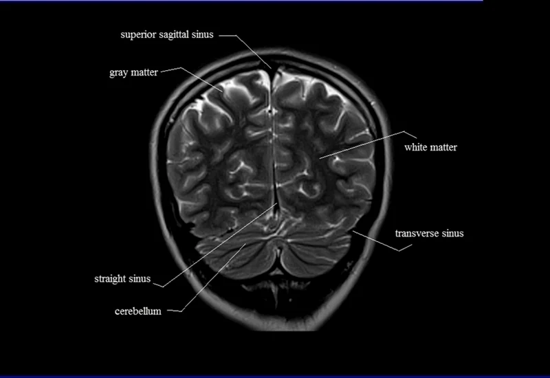
2. Cerebellum
- Located at the back of the brain, the cerebellum is responsible for coordination, balance, and fine motor control.
- MRI can help detect disorders affecting the cerebellum, such as cerebellar atrophy or tumors.
3. Brainstem
- The brainstem controls basic life-sustaining functions like heart rate, breathing, and sleep.
- MRI imaging can reveal structural issues, such as brainstem strokes or infections.
4. Ventricular System
- The brain contains fluid-filled spaces known as ventricles that help cushion and protect the brain.
- MRI allows doctors to examine the size and shape of these ventricles, helping to diagnose conditions like hydrocephalus (excess fluid buildup in the brain).
5. White and Gray Matter
- White matter consists of nerve fibers that transmit signals between different parts of the brain, while gray matter contains the neurons responsible for processing information.
- MRI can show the health of these tissues and identify changes that may signal conditions such as multiple sclerosis or stroke.
Benefits of MRI Brain Anatomy in Diagnosis
MRI brain anatomy provides numerous benefits in diagnosing neurological conditions. These benefits include:
- High-Resolution Imaging: MRI delivers high-quality images that show intricate details of the brain’s structures, helping doctors detect abnormalities that may not be visible with other imaging methods.
- Non-Invasive: Unlike procedures like biopsies, MRI is non-invasive, meaning it doesn’t require surgical intervention. This makes it a safer and more comfortable option for patients.
- No Radiation: Since MRI does not rely on radiation, it can be used multiple times without the risks associated with repeated X-rays or CT scans.
- Early Detection: MRI is particularly useful for identifying early signs of neurological disorders before symptoms become noticeable. Conditions like brain tumors, stroke, and degenerative diseases can be detected in their earliest stages, which improves treatment outcomes.
Common Conditions Detected Using MRI Brain Anatomy
MRI brain anatomy plays a crucial role in detecting a variety of neurological disorders. Some of the most common conditions that MRI can help diagnose include:
- Brain Tumors
- MRI is the primary imaging technique used to detect brain tumors. It provides detailed images that help doctors assess the size, location, and type of tumor.
- Early detection is critical for planning effective treatment, whether through surgery, radiation therapy, or other methods.
- MRI is the primary imaging technique used to detect brain tumors. It provides detailed images that help doctors assess the size, location, and type of tumor.
- Stroke
- MRI is effective in identifying the areas of the brain affected by stroke, whether from ischemia (lack of blood flow) or hemorrhage (bleeding).
- MRI can quickly reveal the extent of brain damage, allowing for immediate intervention to minimize further harm.
- MRI is effective in identifying the areas of the brain affected by stroke, whether from ischemia (lack of blood flow) or hemorrhage (bleeding).
- Multiple Sclerosis (MS)
- MS is a chronic autoimmune disease that affects the central nervous system. MRI scans can detect the lesions or plaques that are characteristic of MS, helping doctors confirm a diagnosis and monitor disease progression.
- MS is a chronic autoimmune disease that affects the central nervous system. MRI scans can detect the lesions or plaques that are characteristic of MS, helping doctors confirm a diagnosis and monitor disease progression.
- Alzheimer’s Disease
- MRI is used to observe brain atrophy, particularly in the hippocampus, a region crucial for memory and learning. This helps in the early diagnosis of Alzheimer’s disease.
- Additionally, MRI is used to track the progression of the disease over time, allowing for adjustments in treatment plans.
- MRI is used to observe brain atrophy, particularly in the hippocampus, a region crucial for memory and learning. This helps in the early diagnosis of Alzheimer’s disease.
- Epilepsy
- MRI can help identify structural changes in the brain that may be causing seizures, such as brain lesions or scarring.
- It is a valuable tool for understanding the underlying cause of epilepsy, which can guide treatment decisions.
- MRI can help identify structural changes in the brain that may be causing seizures, such as brain lesions or scarring.
Functional MRI (fMRI) and Its Role in Brain Activity Mapping
In addition to traditional MRI, Functional MRI (fMRI) has been developed to observe the brain in action. Unlike structural MRI, which focuses on the anatomy of the brain, fMRI measures changes in blood flow within the brain as a result of neural activity. This allows doctors to visualize which areas of the brain are active during specific tasks, such as moving a limb, speaking, or solving a problem.
fMRI is particularly useful in:
- Pre-Surgical Planning: fMRI helps identify critical areas of the brain that control motor functions, language, or vision, allowing surgeons to avoid damaging these areas during surgery.
- Research: fMRI is widely used in research to understand how different regions of the brain interact during cognitive tasks or in response to external stimuli.
Conclusion
MRI brain anatomy is an essential tool for understanding the structure and function of the brain. With its ability to provide highly detailed, non-invasive images, MRI plays a vital role in the early detection, diagnosis, and monitoring of various brain disorders. From brain tumors to degenerative diseases, MRI offers invaluable insights that can lead to more accurate diagnoses and better treatment outcomes. As technology continues to advance, the role of MRI in brain health will only grow, providing even greater precision in the detection and management of neurological conditions.


